Digital Image Analysis in Pathology
DIAPath (Digital Image Analysis in Pathology) is a platform for the validation of tissue-based biomarkers using special staining, standardized immunohistochemistry (IHC) and multiplexed immunofluorescence (mIF), whole slide imaging and image analysis (with artificial intelligence) for objective quantitative analysis.
The overall goal is to extract information useful for understanding disease processes and response to therapy, and to characterize biomarkers useful for diagnostic, prognostic and therapeutic purposes.
Equipments
- Automated Microtome
- IHC/IF/ISH research platform (Dako Autostainer Link 48; Roche Discovery ULTRA)
- Fluorescence whole slide scanner (Zeiss Axioscan 7)
- Brightfield whole slide scanner (Hamamatsu Nanozoomer S360)
- Image processing and analysis (Visiopharm, QuPath, ZEN blue)

Services
- FFPE block manufacturing (Cells, organoid, organs & tissue microarrays)
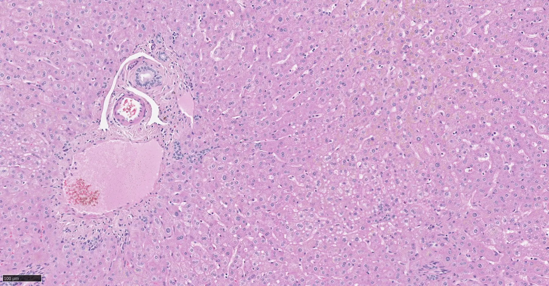
- Histological slide sectioning
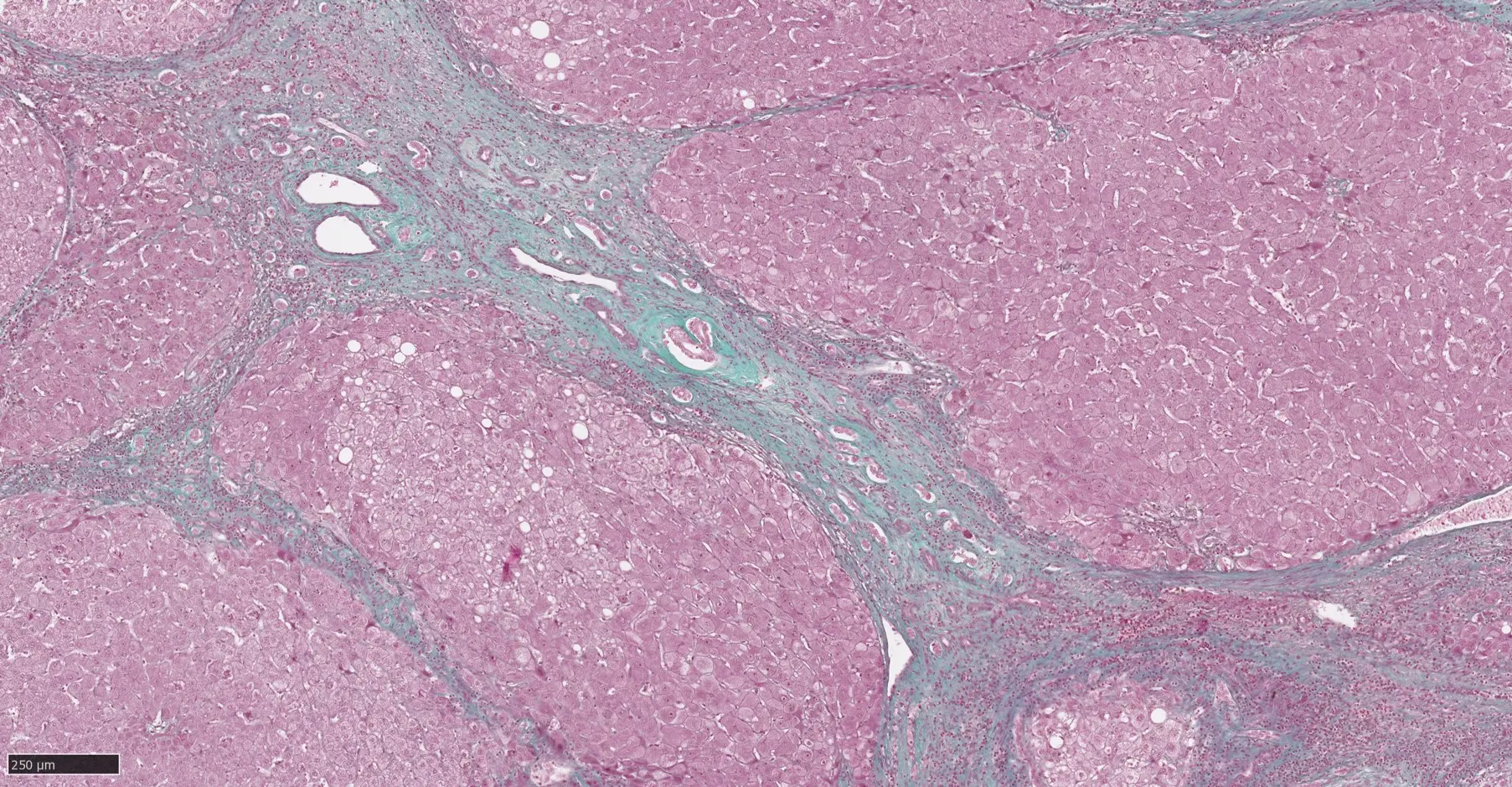
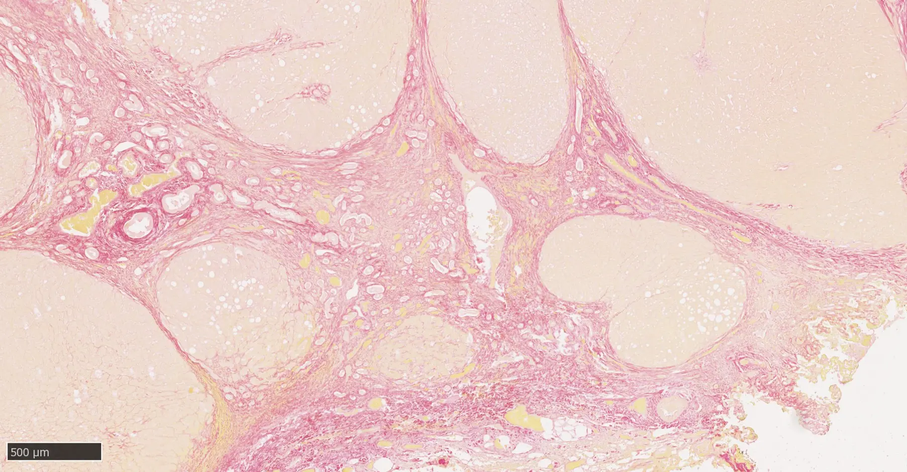
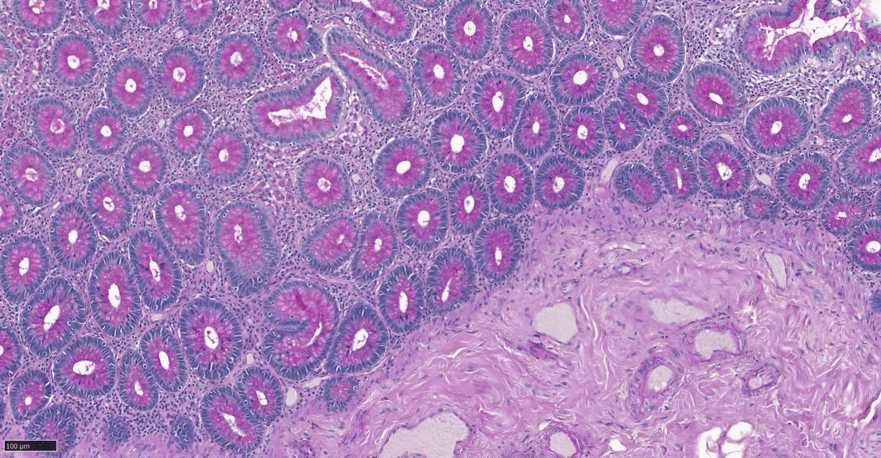
- Special staining
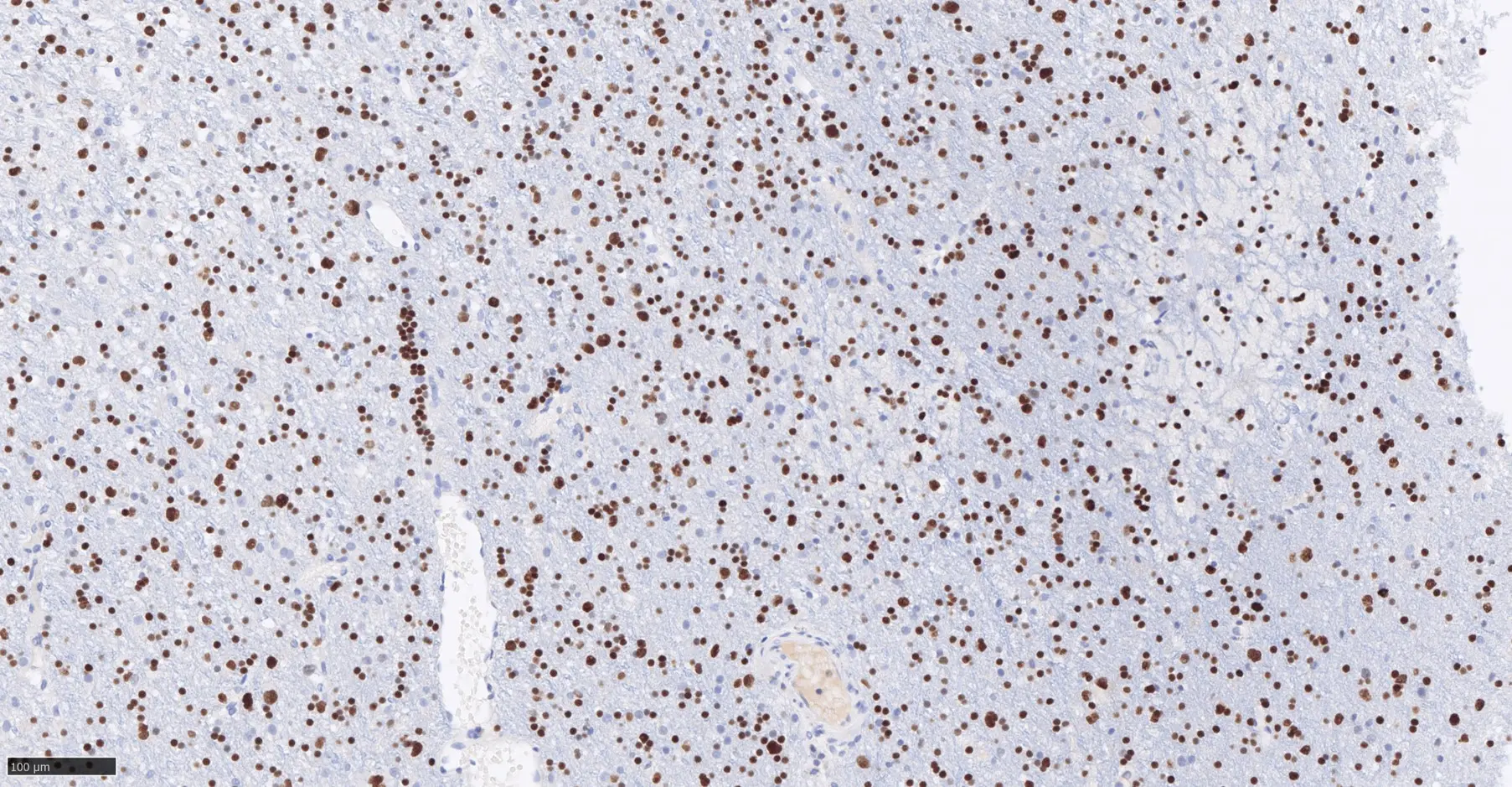
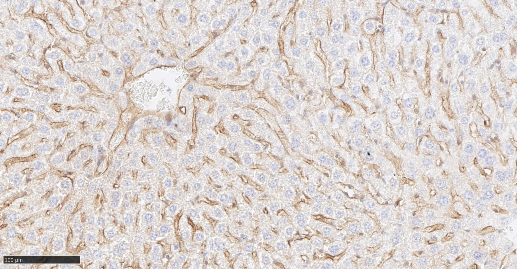
- Standardized immunohistochemistry (List of antibodies - tryptique)
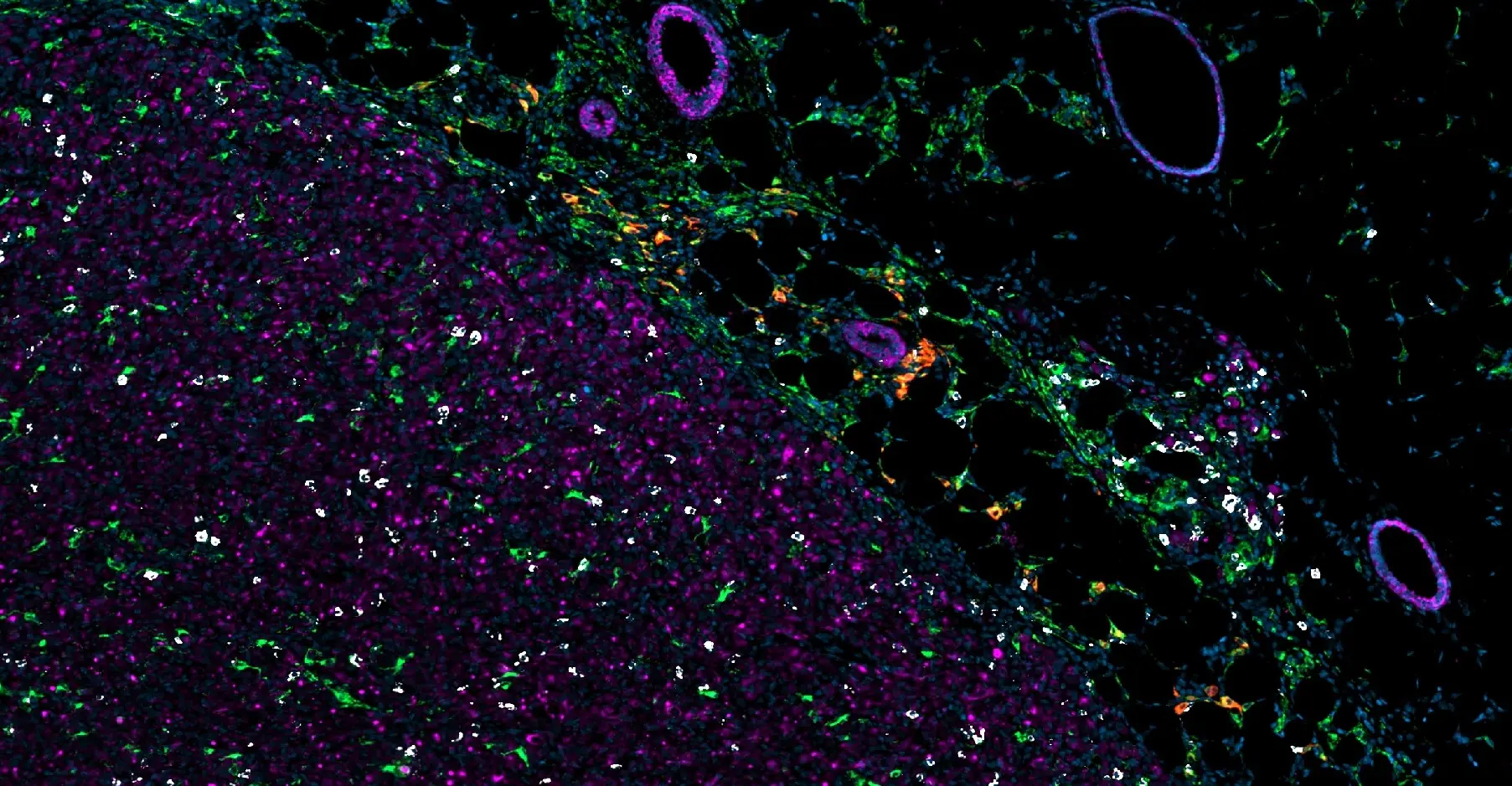
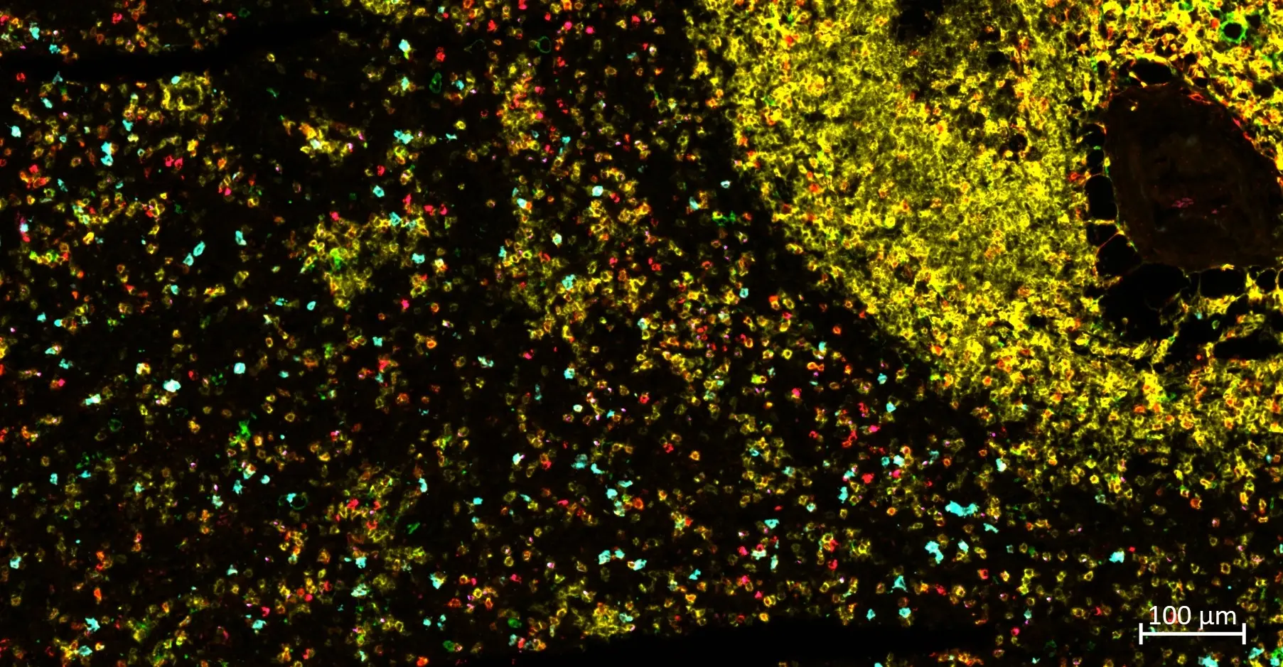
- Multiplexed immunofluorescence
- Chromogenic in situ hybridization
- High resolution slide scanning
- Digital pathology
- Tissue-based biomarker analysis
- Validation of animal models, antibodies and imaging tracers


BOTTIEAU Sophie
Specialized Technologist

ADAMOU YAROU Rabi
Specialized Technologist

GILLARD Chloé
Bioengineer