Microscopy
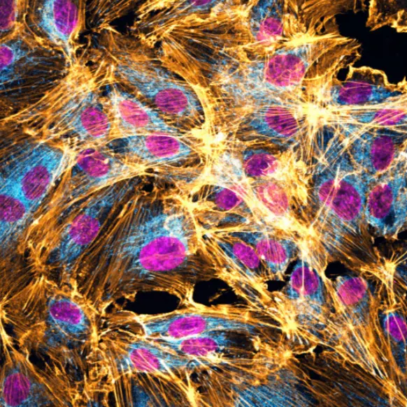
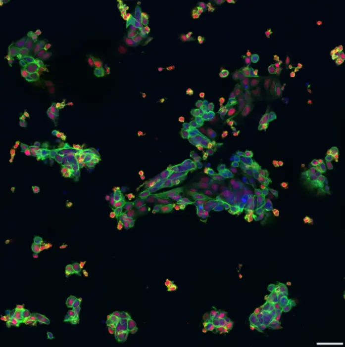
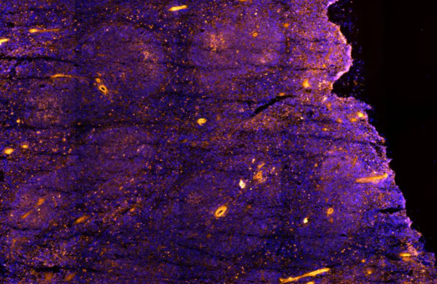
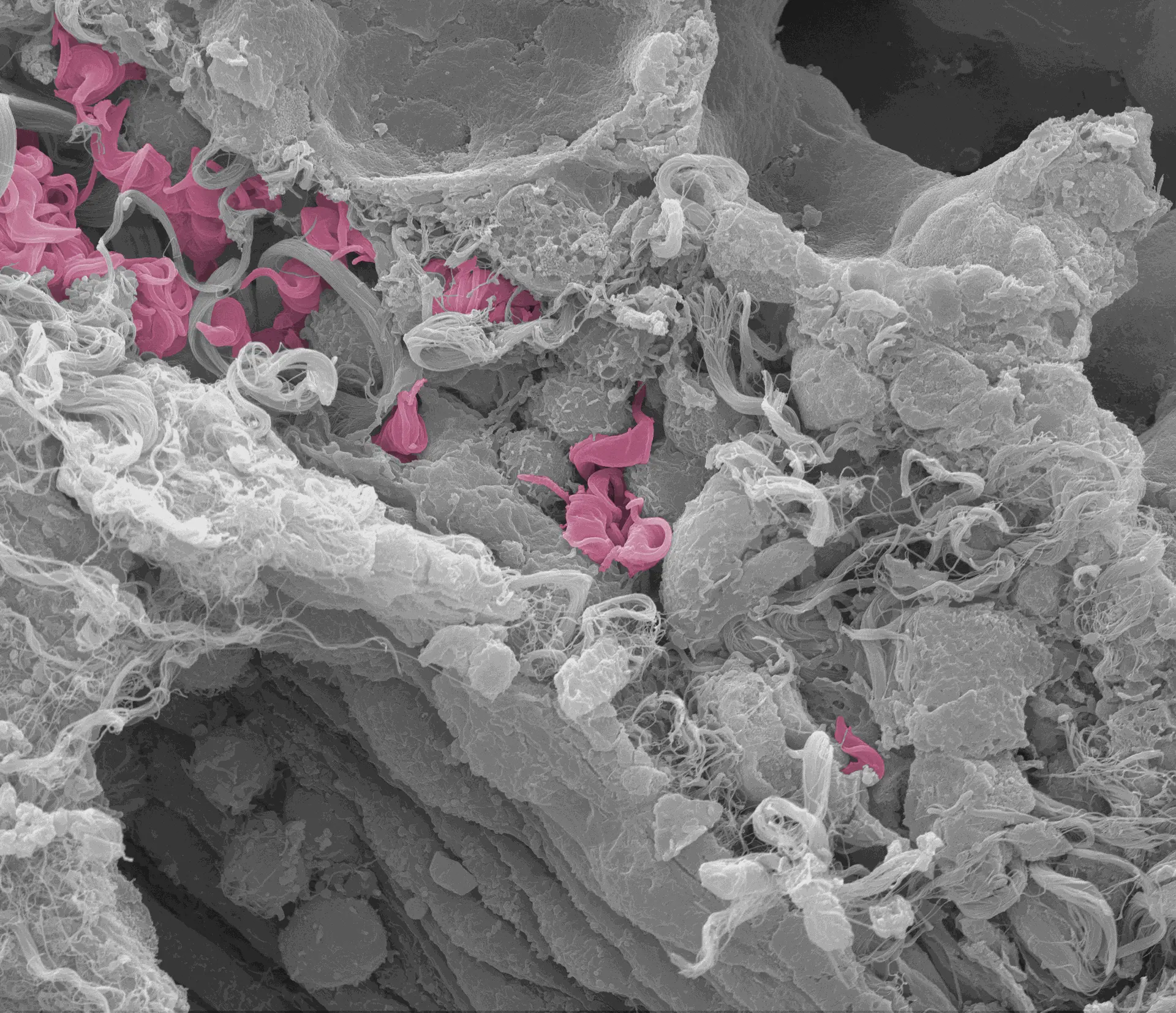
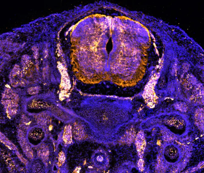
Cellular, sub-cellular and molecular aspects are investigated by using varied cutting-edge microscopy techniques including holography, fluorescence-based imaging and electron microscopy.
Fluorescent and
Holographic Microscopy
Services
- Sample preparation and processing expertise:
- Tissue embedding and sectioning using a cryostat
- Immunofluorescence and other stainings for cells and tissues
- Cell culture and live cell-compatible imaging
- Expansion microscopy protocol development
- Image acquisition:
- Automated high-throughput acquisitions (slide or multi-well plate imaging)
- High-resolution imaging (confocal, super-resolution)
- High temporal resolution (spinning disk, resonant scanner)
- Data interpretation and analysis:
- Qualitative interpretation of images for all types of biological samples, sub-cellular organization interpretation
- Quantitative pipeline development
- Training in fluorescence microscopy techniques
Equipments
- Versatile wide-field fluorescent microscopes
- Confocal microscopes equipped with classic and resonant scanners (Stellaris 5 from Leica), spinning disk confocal microscope
- Super-resolution STED microscope (Stellaris 8 from Leica, equipped with FLIM module)
- FT-IR microscope (Hyperion II from Bruker)


Electron Microscopy
Services
Sample preparation for SEM, TEM, and cryo-EM, and processing expertise:
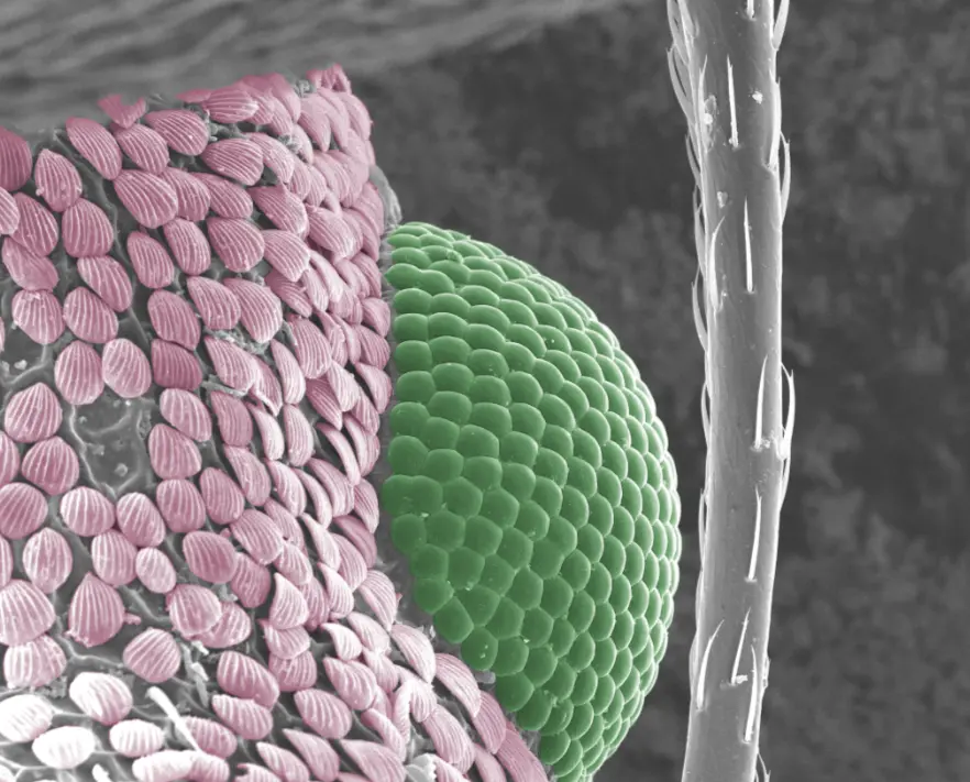
- Sample preparation, sectioning (vibratome), and drying processes for SEM
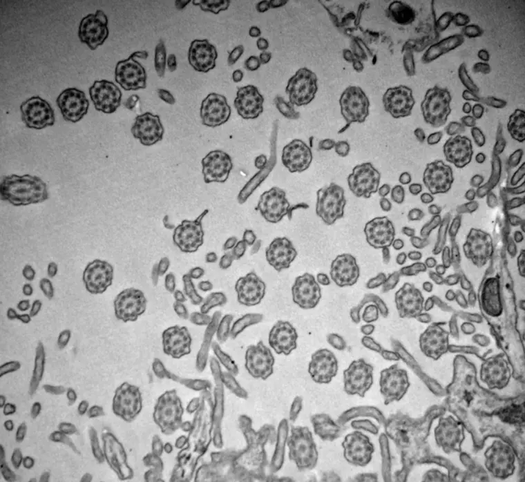
- Cell and tissue embedding in resins for TEM
- Ultramicrotomy
- Tokuyasu technique (immunogold labeling)
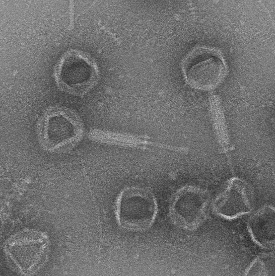
- Negative staining and cryo-plunging (Vitrobot)
Image acquisition:
- SEM imaging at high vacuum or in low vacuum
- 100kV and 200kV RT-TEM imaging
- Single particle analysis (negative staining and in cryogenic conditions)
- RT and Cryo-Electron tomography
- STEM imaging and tomography
Data interpretation and analysis:
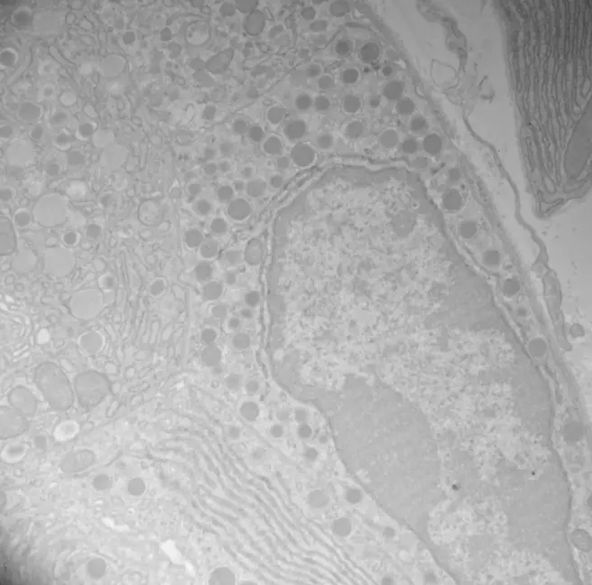
- Ultrastructure detail analysis for all types of biological samples (cells, tissues, etc.)
- Histopathological interpretation
- Single particle structure reconstruction
- Semi-quantitative to quantitative analyses
Training in electron microscopy techniques
Equipments
- Scanning Electron Microscope (SEM - Quanta FEG from FEI)
- Transmission Electron Microscope (TEM - Tecnai 10 100kV from FEI)
- Cryogenic Transmission Electron Microscope (cryo-TEM/Talos FEG 200kV from TFS, equipped with Falcon III camera)
